
 |
Techniques to place intraocular lenses in the absence of adequate capsular support continue to be an important part of the vitreoretinal surgeon’s armamentarium. Shin Yamane, MD, pioneered a modification of sutureless intrascleral IOL fixation using needles to dock and externalize the haptics.1 Here, we describe our approach to optimize the procedure.
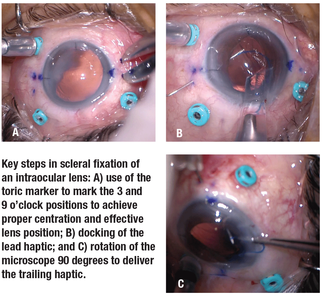
Key steps
• The setup. A toric marker is important to mark the 3 and 9 o’clock positions (Figure A). Calipers are used to mark the conjunctiva 2 mm from the limbus, with another mark placed 2 mm superior (at 3) and 2 mm inferior (at 9). As needed, a standard pars plana vitrectomy and/or lensectomy is performed with cannulas placed away from the above marked locations. A dispersive viscoelastic is placed in the anterior chamber. A keratome fashions a triplanar 3.2-mm clear corneal incision at the superior limbus.
• Lens and injector. The Zeiss CT Lucia 602 three-piece IOL is well suited for intrascleral fixation because of the strength of its haptics. The IOL is loaded in a 3.2-mm Alcon Type A cartridge, delivered through the corneal wound using a Monarch II handpiece (Alcon).
• Delivery. A 0.5-inch long, 30-ga. thin-walled TSK needle is placed on a TB syringe and bent about 75 degrees with the bevel facing inward (docking needle). A standard-bore 30-ga. needle, often found in the operating room, won’t accept most haptics.
The docking needle is inserted with the left hand at 3 o’clock via a tunnel using the previously placed marks. The loaded lens cartridge is inserted into the clear corneal wound with the right hand. The assistant advances the Monarch II injector. The IOL is advanced until the lead haptic emerges (Figure B) and is simultaneously docked into the lumen of the TSK needle before advancing the remainder of the IOL.
Alternatively, the IOL can be delivered partially in the anterior chamber with the trailing haptic outside of the eye and the lead haptic docked into the lumen of the TSK needle using microforceps. The TSK needle is withdrawn and the lead haptic externalized. The haptic is grasped with microforceps and low-temperature cautery thermally deforms the end of the haptic into a nailhead.
Overcoming the challenge
Placing the trailing haptic can be challenging due to awkward hand positions from a standard superior vitreoretinal surgery approach. It becomes ergonomically easier to rotate the seating position and microscope 90 degrees to a nasal approach for right eyes and a temporal approach for left eyes (Figure C, page 14). Again, a 0.5-inch, 30-ga. TSK needle is mounted on a TB syringe and bent almost 90 degrees bevel up. This is inserted along the previous markings at 9 o’clock parallel to the limbus. The needle is brought up to the anterior chamber near the clear corneal wound or even externalized through the clear corneal wound. The second haptic is grasped with microforceps and introduced into the bore of the needle. Once the haptic is docked, the TB syringe is withdrawn, and the externalized trailing haptic is grasped with microforceps and thermally deformed. Both haptics are placed flush with the sclera in the subconjunctival space to center the IOL. RS
 |
REFERENCE
1. Yamane S, Sato S, Maruyama-Inoue M, Kadonosono K. Flanged intrascleral intraocular lens fixation with double-needle technique. Ophthalmology. 2017;124:1136-1142.


