 |
 |
Intravitreal injection remains an efficacious and safe method for drug delivery to the posterior segment for myriad diseases. However, patient and physician treatment burden, as well as the low risk of injection-related adverse effects—e.g., endophthalmitis, retinal tears/detachment and elevated intraocular pressure—remain concerns that may make alternative drug-delivery platforms advantageous.
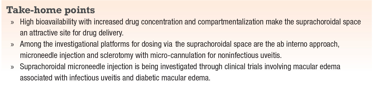
Phase I/II and III trials have demonstrated that using the suprachoroidal space as a site for drug delivery when treating macular edema due to non-infectious uveitis holds promise. With characteristics such as high bioavailability with lower drug doses and compartmentalization of the drug with a potentially favorable side-effect profile (i.e., reduction of IOP-related adverse events), the suprachoroidal space is an attractive site for drug delivery.1,2 Studies have been developed to help optimize this drug delivery platform and evaluate its clinical efficacy.
Although multiple approaches to access the suprachoroidal space exist, including the ab interno approach (Cypass, iStent, etc.) and standard hypodermic needle injection, only a few have demonstrated reliable access to the suprachoroidal space for drug delivery to the posterior segment. Two of these methods are suprachoroidal microneedle injection (Xipere, Clearside Biomedical) and sclerotomy with micro-cannulation (iTrack, Ellex).
Sclerotomy and micro-cannulation
Investigational work has been performed, fashioning a sclerotomy using a specialized microcatheter with a flashing diode to access the suprachoroidal space. One benefit of this platform is that the drug delivery site can be accurately visualized. It has demonstrated promise in animal models as well as in humans as a treatment modality.3-5 Thus far, this technique has been used in humans for treatment of severe subfoveal hard exudates and exudative age-related macular degeneration.3,5
Timothy Olsen, MD, and colleagues have demonstrated that with this approach, medications could be introduced into the suprachoroidal space safely and effectively in porcine and primate subjects.4,7 One shortcoming they described was that suprachoroidal bevacizumab (Avastin, Roche/Genentech) was no longer detected after one week, whereas intravitreal bevacizumab was present 30 to 60 days after injection. Another potential disadvantage of this drug-delivery method is the need for surgery, which may not be convenient for the routine treatment of AMD, uveitis or other conditions where office-based intravitreal injections are the standard-of-care.
The treatment of intraocular tumors has also been considered as a potential application of this microcatheterization technology. Further studies will be needed before it’s widely adopted, but its viability as a surgical procedure has been validated and warrants investigation.
The hollow microneedle
 |
Another promising therapeutic option for suprachoroidal drug delivery is the hollow microneedle. Microneedles consist of a 30-ga. syringe <1 mm in length (usually 900 or 1,000 μm), and are injected posterior to or within the pars plana.6,7 The microneedle is injected in a fashion similar to that of intravitreal injections, and therefore can be utilized in the outpatient setting. Potential drug reflux is generally managed by maintaining the needle within the suprachoroidal space briefly prior to removal.8
Five active clinical trials have been evaluating microneedles for suprachoroidal drug delivery for treatment of noninfectious uveitis and associated macular edema. One of them is the PEACHTREE study, which is investigating the use of Xipere. This trial has shown promising efficacy and safety results.10
Findings from PEACHTREE
In PEACHTREE, a randomized, masked, sham-controlled Phase III trial, 160 patients were enrolled and randomized in a 3:2 ratio to suprachoroid- administered proprietary, preservative-free formulation of triamcinolone acetonide or sham control at baseline and 12 weeks, with primary efficacy and safety endpoints assessed at 24 weeks.
The primary efficacy endpoint of PEACHTREE was a >15-letter gain in best-corrected visual acuity, which 47 percent of patients receiving suprachoroidal corticosteroid achieved vs. only 16 percent of controls (p<0.001).
Patients receiving suprachoroidal corticosteroid injection also experienced an approximately 150-μm reduction of central subfield thickness, compared to 18 μm in controls. IOP elevation rates and cataract progression or development rates were favorable as well compared to controls: IOP-related adverse events were reported in 12 percent of treated patients and 16 percent of controls; and cataract-related adverse events in 7 and 6 percent, respectively.
The Phase II TYBEE trial is investigating Xipere for the treatment of diabetic macular edema. This trial has randomized 71 patients to aflibercept (Eylea, Regeneron) alone or suprachoroidal triamcinolone acetonide with aflibercept. Both arms have shown VA gains (13.5 letters in the aflibercept arm vs. 12.3 letters in the combination arm). Notably, the combination arm required fewer aflibercept treatments (2.8 injections) than the aflibercept-only arm (4.7 injections).
Additional considerations
Lately, there has been research into microneedle injection into the suprachoroidal space with subsequent iontophoresis to drive drug particles to the posterior pole. One study showed a 30 percent increase in drug concentration at the posterior pole after suprachoroidal injection and iontophoresis in an animal model.9 These results may help in the development of supra-
choroidal drug-delivery modalities.
Bottom line
Given its unique tissue distribution, the suprachoroidal space has demonstrated promise as a unique drug-delivery pathway with the
potential to treat inflammatory and vascular diseases of the posterior segment. While both surgical and non-surgical options for accessing the suprachoirdal space have been studied for commonly encountered posterior segment conditions, recent trials have shown the potential of suprachoroidal dosing in the clinic.
Given the favorable drug distribution profile that suprachoroidal drug delivery may offer, its use could offer a multitude of potential benefits, such as a reduction of treatment burden, improved efficacy and safety, and new indications for treatment of posterior segment disease.
REFERENCES
1. Tyagi P, Kadam RS, Kompella UB. Comparison of suprachoroidal drug delivery with subconjunctival and intravitreal routes using noninvasive fluorophotometry. PLoS One. 2012;7:e48188
2. Kim YC, Edelhauser HF, Prausnitz MR. Targeted delivery of antiglaucoma drugs to the supraciliary space using microneedles. Invest Ophthalmol Vis Sci. 2014;55:7387-7397.
3. Rizzo S, Ebert FG, Bartolo ED, et al. Suprachoroidal drug infusion for the treatment of severe subfoveal hard exudates. Retina. 2012;32: 776-784.
4. Olsen TW, Feng X, Wabner K, et al. Cannulation of the suprachoroidal space: A novel drug delivery methodology to the posterior segment. Am J. Ophthalmol. 2006;142:777-787.
5. Tetz M, Rizzo S, Augustin AJ. Safety of submacular suprachoroidal drug administration via a microcatheter: Retrospective analysis of European treatment results. Ophthalmologica. 2012;227:183-189.
6. Goldstein DA. Achieving drug delivery via the suprachoroidal space. Retina Today. 2014;9:82-87.
7. Pearce W, Hsu J, Yeh S. Advances in drug delivery to the posterior segment. Curr Opin Ophthalmol. 2015;26:233-239.
8. Chiang B, Jung JR, Prausnitz MR. The suprachoroidal space as a route of administration to the posterior segment of the eye. Adv Drug Deliv Revi. 2018;126:58-65.
9. Jung J, Chiang B, Grossniklaus H, Prausnitz M. Ocular drug delivery targeted by iontonphoresis in the suprachoroidal space using a microneedle. J Control Release. 2018;277:14-22.
10. CLEARSIDE, XIPERE and PEACHTREE to be front and center at AAO 2018. [press release]. Alpharetta, GA; Clearside Biomedical; October 22, 2018.


