 |
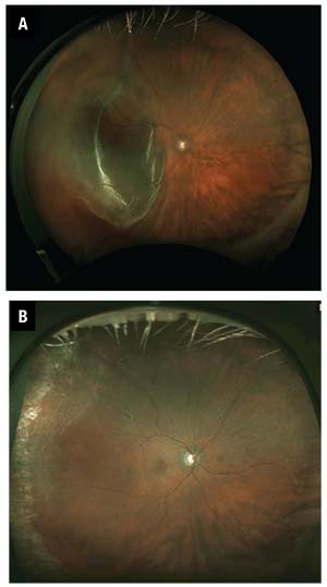
This prompted me to look at my own cases. I reviewed my last 100 cases of repair of retinal detachment (RD) with vitrectomy (CPT code 67108) that had at least six months of follow-up. I excluded cases that involved conditions such as trauma, endophthalmitis or retained lens fragment, or cases that were primarily vitreous hemorrhage with retinal break and a small, localized retinal detachment that might also be coded with 67108, but might have a more favorable prognosis than a bullously detached retina. I also excluded cases that had previous surgeries for RD, such as pneumatic retinopexy and scleral buckling (Figure 1).
I found that I did scleral buckling with pars plana vitrectomy in only 10 cases; I repaired 90 percent with vitrectomy alone. Six of the 100 patients required additional surgeries to repair recurrent retinal detachment, so the one-operation success rate was 94 percent.
Interestingly, all the cases that failed were cases of vitrectomy alone. The one-operation success rate for vitrectomy alone was still a respectable 93 percent (83/90). However, it troubled me that adding the scleral buckling procedure seemed to increase the success rate.
Would my success rate have been higher if I had performed more or all vitrectomies with scleral buckling? Having to incorporate scleral buckling on all my vitrectomy cases was not that appealing to this presbyope.
To Buckle or Not to Buckle
Mark Twain said, “There are three kinds of lies: lies, damned lies and statistics.” So I decided to manipulate my data to improve my vitrectomy-only results. Since I had six failures, I decided to look at my last 100 cases of CPT code 67108 that were vitrectomy only and see if I could raise my success rate to 94 percent by reviewing more cases. I had to review a few more months of surgery.
Unfortunately, I found I had one additional failure with vitrectomy alone, so 93/100, or 93 percent, were repaired with one operation, just as with the 90 cases. However, during that same additional time interval, I did three more vitrectomies with scleral buckling, and two required additional retinal surgeries, resulting in a one-operation success rate of 11/13, or 85 percent.
This was what I’d expected. I usually choose to add scleral buckling on cases that I think may fail from proliferative vitreoretinopathy (PVR) based on three factors:
-how chronic the detachment is;
-the amount of vitreous debris; and
-how rigid the retina is.
Thus, these cases should have had a higher failure rate. These findings were more consistent with my clinical experience that scleral buckling is unnecessary for most primary retinal detachments.
I don’t do all retinal detachment repairs by pars plana vitrectomy. During the time period that I did those 113 operations of retinal detachment repair with vitrectomy, I also performed scleral buckling alone (CPT code 67107) in 43 cases. All but one of those cases required only a single surgery, a 98 percent one-operation success rate.
Overall, my one-operation success for these reviewed cases was 94 percent. Almost two-thirds of the time—64 percent—I performed vitrectomy alone; 28 percent were scleral buckle alone; and 8 percent vitrectomy with scleral buckling.
Based on my small numbers, I do not believe that scleral buckling is superior to vitrectomy, or that scleral buckling with vitrectomy is inferior to vitrectomy alone. Rather, if I choose the appropriate surgery to repair a retinal detachment, the single operation success rate is very high—more than 90 percent. So, how do I decide on the appropriate surgery?
When To Do Scleral Buckling
As a general rule, if the retinal detachment is not due to posterior vitreous detachment, I will perform scleral buckling. These cases are often due to high myopia, abnormal retinas with holes or lattice, and have abnormally
 |
| Figure 1. A typical case that would be included in the CPT code 67108 series, retinal detachment (RD) repair with vitrectomy (A). Postoperative photography (B) shows outcome of macula-off RD, resulting in complete Recovery of central vision and subjectively better vision post-RD repair because of vitrectomy and floater removal. |
Some of the most spectacular retinal detachment failures that I have seen are cases where vitrectomy alone was performed for a young myope and the patient ends up with PVR and multiple surgeries with a horrible visual outcome. The retina doesn’t conform to the shape of the eyeball, which is why it detached in the first place, and removing the vitreous (assuming that the vitreous is removed with successful separation of the posterior hyaloid, which is not always the case) and lasering retinal holes may not be adequate to permanently reattach the retina. Sometimes it takes three or four operations and a large relaxing retinectomy to successfully reattach the retina, but by this point the patient has usually lost vision.
For retinal detachments that are due to vitreous detachment with concomitant tearing of the retina, the obvious choice is vitrectomy, especially if vitreous hemorrhage, significant vitreous pigment and haze, or symptomatic floaters are present. Except when pneumatic retinopexy is appropriate, vitrectomy is the correct procedure, and that is what I do in almost all pseudophakic patients.
In instances of retinal detachment due to PVD where the vitreous and lens are clear, or in situations such as when the patient has a need to travel, I will sometimes perform scleral buckling surgery. Of the 43 cases of scleral buckling alone (CPT code 67107) that I did during the review period, all were phakic; half (23/43) had PVD causing the retinal detachment.
Besides the cases in which I perform vitrectomy with scleral buckling that I mentioned earlier, I’m also more likely to perform scleralbuckling with vitrectomy in younger patients and those who are phakic because my vitrectomy may not be as complete. The location of retinal break(s) does not factor into my decision to incorporate scleral buckling.
My Approach To Scleral Buckling
As someone who trained in the late 1980s and early 1990s, I used radial and segmental buckling when I first started out in practice. I have not used a radial element in nearly 20 years. A gas bubble or vitrectomy will prevent “fish-mouthing” of a retinal tear. I rarely do segmental buckles, except
 |
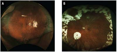
| Figure 2. A poorly performed drainage retinotomy (A) is too posterior and too large. Despite single-operation success, the patient is unhappy because of a visual scotoma. This retinotomy (B) is not as symptomatic, and although the scotoma is apparent to the patient, it is not as bothersome as that in Figure 2A because it is farther from optic nerve and is in the superior field of view, which is often less noticeable. However, it is still unnecessarily large. |
I perform external drainage on almost all cases of retinal detachment repair without vitrectomy (41/43). I use a short, 25-gauge needle for drainage, for which I sometimes use indirect ophthalmoscopic visualization (Figure 2). Rarely I will perform a posterior sclerotomy and suture the sclerotomy, usually on chronic detachments when the subretinal fluid is very thick.
My Approach to Vitrectomy
I perform almost all vitrectomies for retinal detachment with a posterior retinotomy and internal drainage of subretinal fluid. When I drain through a peripheral break, my removal of subretinal fluid is less complete. This has sometimes resulted in subretinal fluid being pushed into the macula, when the macula was not involved, and, less frequently, a macular fold. I use C3F8 on almost all cases, phakic or pseudophakic.
 |
| Figure 3. Retinotomies should be like these three, as highlighted with arrows: small and visually insignificant. |
I am not concerned about causing cataract; I use a longer-acting gas, because cataract surgery is very successful and safe today and because retinal detachment is blinding if my surgery fails.
I occasionally use SF6 or silicone oil if the patient asks for it because of travel needs. I rarely use perfluorocarbon liquid unless a giant retinal tear is involved or if the patient is young with an abnormally adherent vitreoretinal interface and I need it to stabilize the retina to adequately remove the vitreous safely.
My usual sequence for reattaching the retina involves flattening the retina by removing the fluid from the vitreous cavity and then draining through the posterior retinotomy. I then perform laser to the peripheral retina. After completion of the peripheral laser, I drain the fluid that has migrated posteriorly from the vitreous base at the optic nerve.
Lastly, I drain again from the posterior retinotomy and then apply the laser to it. I find that if I perform laser to the posterior retinotomy immediately after the initial internal drainage, the fluid that migrates posteriorly while I am doing the peripheral laser lifts up the edges of the retinotomy and I can no longer see my laser burn. This results in my treating the retinotomy again and creating a larger laser scar than desired.
| Take-home Point Surgery to repair retinal detachments has improved dramatically over the past three decades. With improvements in the instrumentation for vitrectomy surgery, such as wide-angle viewing, illuminated lasers and small-gauge/high-speed vitrectomy cutters, and the more frequent use of vitrectomy for retinal detachment repair, we can confidently tell patients that we have a better than 90-percent chance of repairing their detachment with one operation and restoring their vision. These advances in retinal detachment surgery, in the author’s opinion, are almost as great as the improvements we have seen in the treatment of macular diseases with anti-VEGF medications. |
I try to make my posterior retinotomies not too posterior—usually several disc diameters from the optic nerve and arcades—to reduce the risk of visual scotomas; and I try to keep the size under 1 disc diameter (Figure 3).
I have heard speakers advocate not applying laser to the retinotomy, and instead making a more posterior retinotomy near the nerve and around the arcade, presumably to make subretinal fluid drainage more complete. I have never personally done that.
For laser retinopexy, I do not use continuous laser because I have very slow reflexes; by the time I see that the retina has turned white, I have probably “overcooked” it. I have seen a trend of patients having one row of heavy continuous laser in the peripheral retina around or just anterior to the equator. Typically I see them as a second opinion when they develop breaks on either side of the laser. I am not sure when this type of laser treatment became fashionable since I trained a long time ago. Truthfully, I would have never thought to perform laser in this fashion as I fail to see the rationale behind it.
In the areas in which I perform laser, I treat the retina the same way I was taught to treat retinal breaks in the clinic—to the ora serrata.
I use a curved illuminated laser and perform scleral depression myself to get treatment all the way to the ora serrata without damaging the lens in phakic eyes. I try to obtain a light gray burn to create a chorioretinal scar but without coagulation or shrinkage of the retina. I use a more intense white burn when I have performed a relaxing retinectomy and I’m not concerned about the retina contracting from the laser because there is a free edge.
Lastly, I suture my sclerotomies in cases when I want a more complete gas fill, such as inferior detachments and cases with early PVR and all cases involving silicone oil or scleral buckling. RS


