 |
|
Bio Dr. Abbey is director of clinical research at Texas Retina Associates, Dallas, and a clinical assistant professor of ophthalmology at the University of Texas DISCLOSURES: Dr. Abbey is a consultant to Alcon, Allergan/AbbVie, Alimera Sciences, EyePoint |
The 40th annual scientific meeting of the American Society of Retina Specialists convened in New York City in July, bringing together retina specialists from around the world with a return to a full live format.
Among the cutting-edge clinical science presented at the meeting, here we review five abstracts that are worth a second look:
• A multicenter trial of adults with rhegmatogenous retinal detachment treated with a scleral buckle or combined pars plana vitrectomy-SB procedure.
• A hospital-based, prospective, randomized study of eyes with large macular holes treated with either the multilayered inverted limiting membrane flap technique or internal limiting membrane peel.
• Experience with a point-of-care program with optical coherence tomography for people with central retinal artery occlusion.
• A Phase II trial of an investigational antisense oligonucleotide for geographic atrophy.
• Results of a Phase III trial of an agent to reverse pharmacologically induced mydriasis.
Repair techniques for rhegmatogenous retinal detachment
 |
This multicenter, prospective, nonrandomized comparative trial enrolled 83 adult patients with a primary macula-off rhegmatogenous retinal detachment who had scleral buckle or a combined pars plana vitrectomy-SB procedure. The goal was to evaluate the risk of low-integrity retinal attachment (LIRA), characterized by retinal displacement on fundus autofluorescence.1
In presenting the results, Aditya Bansal, MD, a vitreoretinal clinical fellow at St. Michael’s Hospital in Toronto, said recent evidence suggests that LIRA is more likely to occur with PPV compared with a retinal pigment epithelium-pump procedure such as SB or pneumatic retinopexy—most likely due to the full-gas fill used during PPV that exerts a buoyant force on the retina and residual subretinal fluid, leading to stretching of the retina and worse function outcomes.
All patients had FAF and functional outcomes assessment. Two masked graders assessed the FAF images for retinal vessel printings and adjudicated any differences by consensus. Excluded were patients with poor-quality ungradable FAF images. Primary outcome was the risk of LIRA following SB vs. PPV-SB.
The study found that SB is associated with less retinal displacement/LIRA than PPV-SB, although functional outcomes were similar between the two groups.
Of the 83 eyes in the study, 41 percent (n=34) had SB and 59 percent (n=49) had PPV-SB. Two graders evaluated the FAF findings with a high rate of agreement in detecting LIRA (85.5 percent, Kappa=0.629; 95% CI=0.439–0.819).
Baseline characteristics differed with younger phakic patients in the SB group and patients with more extensive detachments in the PPV-SB group. In the SB group, 17.6 percent (n=6) had LIRA vs. 38.8 percent (n=19) in the PPV-SB group (p=0.039).
The study found a statistically significant association between LIRA and procedure type remained after a multivariable logistic regression adjusting for gender, lens status, extent of retinal detachment in quadrants and baseline logMAR visual acuity with odds of retinal displacement of 3.875 (95% CI=1.18–12.723, p=0.026) for PPV-SB vs. SB. There were no statistically significant differences in functional outcomes between the two groups.
Dr. Bansal said that further prospective studies with a larger sample size are required to assess the impact of retinal displacement on functional outcomes in patients undergoing SB or PPV-SB.
Dr. Bansal had no relevant financial relationships to disclose.
Comparison of repair techniques for large macular holes
 |
Much controversy surrounds the use of two techniques for repairing large macular holes: the multilayered inverted internal limiting membrane (ML-IILM) flap technique with perfluorocarbon liquid; and the standard internal limiting membrane peeling technique.
Vishal Agrawal, MD, a professor at SMS Medical College in Jaipur, India, reported on a hospital-based, prospective, randomized, interventional consecutive study of 150 eyes, with an even number assigned to either the ILM peeling group or vitrectomy with the ML-IILM technique.2 Patients were treatment-naive, age 50 years or older and had a full-traction macular hole with a base diameter of ≥600 µm. Excluded were patients with amblyopia, inflammatory eye disease, hypertensive and diabetic retinopathy, retinal detachment or retinal surgery, glaucoma, ≥−6D myopia or if they refused consent.
 |
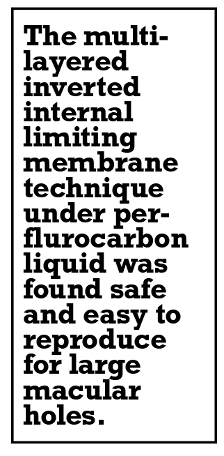
The study found that ML-IILM had significantly better rates of anatomical closure and visual outcome. Final follow-up was at one year. During follow-up at one, three, six and 12 months, the mean postoperative best-corrected visual acuity was significantly better in the ML-IILM group: 0.12 ±0.07 vs. 0.20 ±0.11; 0.14 ±0.10 vs. 0.22 ±0.13; 0.18 ±0.11 vs. 0.30 ±0.12; and 0.19 ±0.12 vs. 0.31 ±0.14, respectively (p=0.001 for all). Anatomical closure was achieved in all patients who had ML-IILM and 93.3 percent (n=70) in the ILM group.
Type 1 closure rates were also better in the ML-IILM group: 93.3 percent (n=70) vs. 78.7 percent (n=59). Type II closure occurred in 6.7 percent (n=5) of the patients who had ML-IILM vs. 15.6 percent (n=11) (p<0.05). Among the ILM patients, 6.67 percent (n=5) of cases failed to achieve any anatomical closure.
Dr. Agrawal said the ML-IILM technique under PFCL is a safe and easily reproducible and offers distinct advantages over ILM peeling in terms of flap-related complications.
Dr. Agrawal and colleagues had no relevant financial relationships to disclose.
Point-of-care OCT and management of central retinal artery occlusion
 |
Central retinal artery occlusion can be visually debilitating and there’s no universally accepted treatment. Fibrinolysis has been shown to improve visual outcomes, but timing is critical. Patients must have treatment within hours of symptom onset, but diagnosis delays reduce chances to improve their vision.
For this study, optical coherence tomography machines were installed at three stroke centers in the Mount Sinai health system in New York City.3 The goal was to evaluate whether point-of-care diagnosis can improve vision and functional outcomes. Gareth Lema, MD, PhD, of New York Eye and Ear of Mount Sinai, reported that the point-of-care OCT in stroke centers can expedite time to treatment for CRAO, increase eligibility for patients to receive treatment and maximize visual improvement.
 |
Dr. Lema noted that any patient who reports to the emergency department with painless monocular vision loss suspicious for a CRAO or stroke activates the stroke protocol. Simultaneously, the retina service gets an alert that a possible CRAO has arrived. The stroke team evaluates the patient, including visual acuity data, pupil exam and OCT of the macula. The clinical data and images are transmitted to members of the retina service who assist in making the diagnosis.
If they confirm a CRAO and the patient can be treated within 12 hours of stroke onset, the patient goes directly for treatment with intra-arterial recombinant tissue plasminogen activator (tPA). If another diagnosis is considered at any time, a full ophthalmology consult is performed before any treatment is considered. Patients who aren’t eligible for treatment still get evaluated with a full stroke work-up. Upon admission, patients are followed by the ophthalmology service to document visual outcomes and safety.
Dr. Lema reported on the experience with the protocol after one year. The study included 35 patients, 19 of whom had CRAOs. Seven of eight patients who met the treatment criteria for intra-arterial tPA (17 mg) received the treatment. All other patients underwent the stroke work-up.
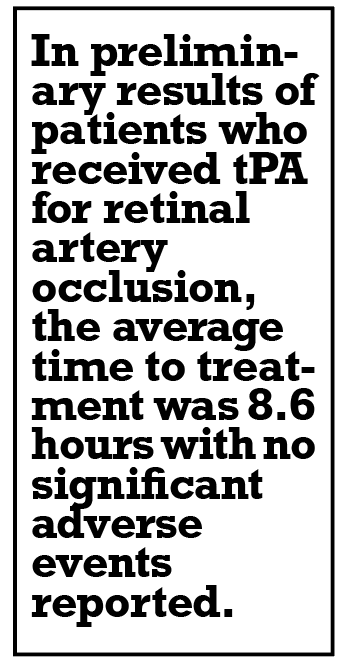
In the preliminary results of five patients who received tPA, the average time to treatment was 8.6 hours (range: 6.5 to 11.75 hours). Visual gains were measured within 24 hours of treatment and all patients showed improvement in visual acuity. Interestingly, two patients lost vision again within two days of treatment, presumably from secondary emboli or thrombosis. In one, antiplatelet therapy reversed the vision loss. No significant adverse events were reported in patients who received tPA.
Dr. Lema had no relevant financial relationships to disclose.
Baseline characteristics for Phase II trial of antisense therapy for GA
 |
IONIS-FB-LRx (Ionis Pharmaceuticals) is a novel investigational antisense oligonucleotide targeting liver factor B in the complement cascade as a treatment for geographic atrophy. Glenn Jaffe, MD, of Duke University, reported on the baseline characteristic for the ongoing Phase II GOLDEN trial,4 the goal of which was to determine whether treatment with IONIS-FB-LRx reduced GA growth in eyes with age-related macular degeneration.
Dr. Jaffe noted the Phase I data supported further development of IONIS-FB-LRx. Based on analysis of the first 100 subjects in the Phase II trial, the median participant age is 76; approximately 58 percent are female. Two-thirds of the lesions are subfoveal in nature, 65 percent are multifocal, 95 percent are bilateral and the mean baseline GA area is 7.6 mm2.
The mean annualized GA area change is approximately 2 mm2 during the screening period. The growth rate was fastest for nonfoveal-centered multifocal lesions and slowest for unifocal nonfoveal-centered lesions. The baseline BCVA was more than 2 lines better for multifocal lesions than unifocal lesions and worse for foveal-centered lesions on average.
Dr. Jaffe said the baseline characteristics and annualized growth rates were similar to previously reported studies of investigative agents for GA, including the CHROMA/-SPECTRI trials of lampalizumab, the FILLY trial of pegcetacoplan and the GATHER 1 trial of avacincaptad pegol.
Dr. Jaffe disclosed financial relationships with Novartis, Regeneron Pharmaceuticals and Roche.
Phase III results of phentolamine to reverse mydriasis
 |
Phentolamine ophthalmic solution (POS) 0.75% is designed to reverse pharmacologically induced mydriasis. David Boyer, MD, of Retina-Vitreous Associates Medical Group in Los Angeles, reported that the MIRA-2 and MIRA-3 Phase III trials have shown that POS reduced mydriasis within 60 to 90 minutes in most patients.5
MIRA-2 and MIRA-3 were multicenter, randomized, placebo-controlled, double-masked clinical trials in healthy subjects (n=553; age ≥12 years). Stratified by iris color, subjects were randomized to mydriatic agent 3:1:1 (2.5% phenylephrine, 1% tropicamide or Paremyd, respectively) and treatment 1:1 in MIRA-2 and 2:1 in MIRA-3 of POS or placebo. The primary endpoint was percent of subjects returning to ≤ 0.2 mm from baseline photopic pupil diameter (PD) at 90 minutes. Secondary endpoints included time to return to baseline PD, visual acuity, time savings and change from maximum dilation.
Across both studies, 338 patients received POS (average age 33 years, 60 percent female) and 215 received placebo (average age 33 years, 62 percent female). Across all mydriatic agents at 90 minutes, 49 percent and 58 percent of study eyes in MIRA-2 and MIRA-3, respectively, that received two drops of POS returned to ≤0.2 mm of baseline compared to 7 percent of placebo-treated subjects in MIRA-2 and 6 percent in MIRA-3 (p<0.0001).
Similar efficacy was also seen starting at 60 minutes and lasting up to 24 hours (p<0.0001) with one or two drops of treatment and across light and dark irides. Mean pupil diameter was significantly lower with POS at all time points starting at 60 minutes (p<0.0001).
POS was also found to produce a time savings of three to four hours. Adverse events were observed in around 5 percent of patients. They included mild, transient conjunctival hyperemia (11.5 percent, n=39) in POS patients and mild installation site discomfort. No serious AEs were reported across both trials and no patients withdrew from the studies because of them. Additionally, POS didn’t compromise distance visual acuity.
Dr. Boyer disclosed being a consultant to Ocuphire Pharmaceuticals, sponsor of the studies. RS
REFERENCES
1. Bansal A, Kohler J, Ryan E, et al Retinal displacement after scleral buckle vs combined scleral buckle and vitrectomy for the management of rhegmatogenous retinal detachment: ALIGN SB vs PPV-SB. Paper presented at the American Society of Retina Specialists annual meeting; July 14, 2022; New York, NY.
2. Agrawal V, Gupta V, Gupta A. Multilayered inverted internal limiting membrane flap technique vs standard internal limiting membrane peeling for large macular holes: comparative study. Paper presented at the American Society of Retina Specialists annual meeting; July 14, 2022; New York, NY.
3. Lema G. Point-of-care diagnosis of CRAO with OCT at the stroke center: Lessons learned in our first year. Paper presented at the American Society of Retina Specialists annual meeting; July 16, 2022; New York, NY.
4. Jaffe G, Yang Q, Barrett T, et al. Baseline characteristics in the Phase 2 GOLDEN study of IONIS-FB-LRx, an investigational antisense oligonucleotide designed to treat AMD-associated geographic atrophy. Paper presented at the American Society of Retina Specialists annual meeting; July 16, 2022; New York, NY.
5. Boyer D, Patel R, Brigell M, et al. Phentolamine ophthalmic solution for mydriasis reversal, results of the MIRA trials. Paper presented at the American Society of Retina Specialists annual meeting; July 16, 2022; New York, NY.


