
 |
The NGENUITY 3D system for vitreoretinal surgery (Alcon, Fort Worth, Texas), consists of a 4,000-pixel display with an ultrafast, high-definition digital camera that allows for high-resolution, three-dimensional viewing in the operating room. At Associated Retinal Consultants in Royal Oak, Mich., Bozho Todorich, MD, PhD, Aristomenis Thanos, MD, Alan J. Ruby, MD, and George A. Williams, MD, have used this system for over six months and have transitioned to its exclusive use for vitreoretinal cases. In this pearl they share the advantages and limitations of this device for routine use. (Click here to view the video.)
Four Potential Benefits
In the operating room, here is how NGENUITY 3D performs in these key areas:
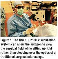
• Surgeon ergonomics. An American Academy of Ophthalmology survey noted high prevalence of neck, upper- and lower-extremity pain in 52 percent of surveyed physicians. Operating with NGENUITY has allowed a more ergonomic posture during surgery (Figure 1), resulting in noticeably less back pain and fatigue at the end of longer operating days.
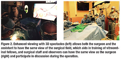
• Assistant visualization. The traditional assistant scope commonly provides a view inferior to the surgeon’s scope. The NGENUITY 3D display eliminates this disparity, offering the same view to the surgeon and the assistant at all times. This enhances the surgeon’s ability to direct and teach surgical staff and trainees, and improves the quality of recorded surgical videos (Figure 2).
• View and stereopsis. NGENUITY 3D augments depth perception, which is particularly helpful during macular surgery and difficult diabetic dissections where axial identification of fine planes is paramount. The NGENUITY 3D camera can also digitally amplify the recorded signal to reduce endoillumination while maintaining sufficient visualization. Decreased endoillumination during macular cases may reduce the risk for photoxicity and also minimizes glare during the fluid-air exchange.
 |
• Intraoperative imaging integration. NGENUITY 3D has the ability to project preoperative imaging such as optical coherence tomography scans and fluorescein angiograms on the display screen during any portion of the operation. This creates a digital platform to enable multimodal surgeon interaction and, possibly, future integration with intraoperative OCT technology, among other potential capabilities.
Potential Limitations
Some limitations of this technology include the cost of acquisition and initial learning curve. Although the surgeons at Associated Retinal Consultants found the adaptation for posterior segment surgery to be rapid, the hyperstereo of the digital camera made anterior segment surgery and external maneuvers, such as conjunctival closure, initially more difficult.
Also, the ideal position for the display screen is directly in front of the surgeon, but this forces the assistant surgeon to turn his or her head 90 degrees, which may make assisting with surgical maneuvers more difficult.
| View the Video Watch Drs. Todorich and Williams perform primary and complex vitreoretinal surgical maneuvers including macular cases, diabetic total-retinal detachment dissections and a complex proliferative vitreoretinopathy case using the NGENUITY 3D viewing system. Click here to view the video. |
Dr. Hahn is an associate at New Jersey Retina in Teaneck. Drs. Todorich and Thanos are second-year vitreoretinal surgery fellows at Associated Retinal Consultants and Oakland University William Beaumont School of Medicine, Rochester, Mich. Drs. Ruby and Williams are partners at Associated Retinal Consultants/Oakland University William Beaumont School of Medicine, where Dr. Williams is chair of the Department of Ophthalmology.
DISCLOSURES: Dr. Williams is a consultant for Alcon. The other co-authors had no conflicts to disclose.


