| Watch the Video A video that describes postoperative macular fold repair and positioning afterward is available at: http://goo.gl/wXXI6c |
When and Why Folds Happen
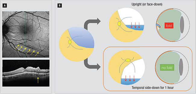
These folds typically occur when enough residual subretinal fluid is focally sequestered, creating redundant retina that folds over itself under intraocular tamponade as the fluid is resorbed. Unexpected folds usually arise with bullous superior retinal detachments and are particularly problematic when the inferior edge crosses the macula.
Why? Because of gravity. Fluid settles beneath the bubble and pools at the inferior edge, which is always the location of the postoperative fold. Folds generally don’t occur in inferior detachments or total detachments.
Often surgeons position a patient upright for tamponade of the retinal break causing a superior detachment. This has great potential to induce a fold under the previously described circumstances. If there is residual subretinal fluid and concern for a fold, many retina specialists may recommend face-down positioning.
 |
| Postoperative fold inferior to the fovea following superior retinal detachment repair (A) is easily seen with autofluorescence and OCT imaging (yellow arrows). Although small and extrafoveal, this fold caused significant distortion and visual rotation. Following vitrectomy for repair of bullous superior retinal detachments (B), upright (or face-down if the detachment extends beyond the fovea) positioning forces residual fluid to gravitate to the inferior edge of the detachment and can promote a fold. Temporal side-down positioning for the first hour immediately postoperatively shifts fluid away from the macula to prevent fold formation. |
However, face-down positioning is often physically difficult for patients and thus not strictly performed, especially as the patient is maneuvered in the postanesthesia care unit (PACU).
The time immediately following surgery is critical in determining the location of fluid sequestration as the retinal pigment epithelium pumps start reapposing the retina. If the patient is only partially face down during this crucial period, fluid will still be sequestered at that inferior edge and a fold may result. And even when face-down positioning is done properly under the best of circumstances, fluid can still pool if the detachment extends beyond the fovea or the apex of face-down positioning.
The Case for Temporal Positioning
Cynthia Toth, MD, a pediatric retinal specialist and retinal surgeon at Duke Health in Durham, N.C., teaches a valuable pearl with regard to patient positioning. As a pioneer of macular translocation surgery in which retinal folds and/or rotations are induced intentionally, she is well-versed in the pathogenesis of these folds.
Dr. Toth recommends first positioning the patient to lie temporal side-down for one hour. She recommends this positioning immediately at the conclusion of surgery, even before the patient is wheeled out of the operating room and into the PACU, to avoid upright positioning. The patient finds this easier than face-down positioning.
After one hour, the patient can be discharged from the PACU with standard positioning instructions to tamponade the retinal breaks (typically upright for a superior detachment). Immediate temporal side-down positioning allows sequestered fluid to shift away from the macula to the periphery, and that one hour is sufficient to allow the RPE pumps to start reapposing the macula, preventing fluid reaccumulation in the macula. In theory, a fold may still occur in the periphery, but this would likely not be visually significant and fortunately does not typically occur.
The purpose of this positioning is not to drain fluid from the retinal break but rather to shift residual fluid away from the macula as the retina is reattaching and avoid the “downward sag” of the retina. Of course, removing all subretinal fluid will also prevent folds.
But for superior detachments with any residual fluid or an inadequate view at the end of surgery to assess for residual fluid, remember this: temporal-side down for the first hour. RS
Dr. Hahn is an associate at New Jersey Retina in Teaneck. Disclosures: Dr. Hahn serves as a consultant for Second Sight Medical Products and Bausch + Lomb.


