A large traumatic macular hole poses a challenge to the vitreoretinal surgeon, because both characteristics—large and traumatic—are associated with lower surgical closure rates. The mechanism of traumatic macular hole formation is more likely to result in avulsed tissue, and even after vitrectomy with wide internal limiting membrane peel, the hole may not successfully close.
 |
 |
Human amniotic membrane scaffold
Newer techniques have been developed to address macular holes with lower likelihood of closure. Stanislao Rizzo, MD, and colleagues at the University of Florence in Italy recently reported on a technique that uses human amniotic membrane (HAM) placed in the subretinal space to bridge the tissue gap and provide a scaffold which the surrounding retinal tissue can overfill to successfully close the macular hole.1
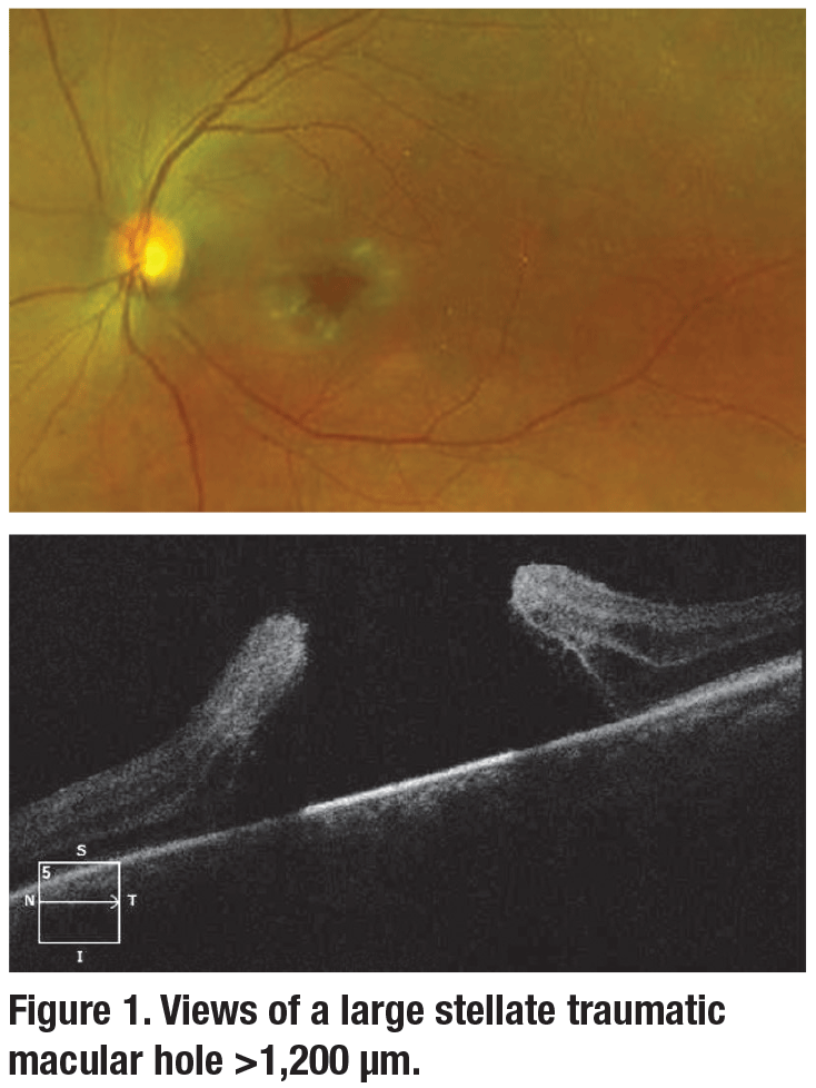
At the University of Colorado, this technique was employed in the repair of a >1,200 μm traumatic macular hole with a stellate configuration (Figure 1). A traditional 25-gauge vitrectomy setup was employed with indocyanine-green-assisted ILM peeling. A multi-segment centripetal ILM peel was performed due to the stellate nature of the traumatic macular hole, paying careful attention to avoid radialization of the leaflets or enlarging of the hole.
The HAM utilized was AmnioGraft (Bio-Tissue), a cryopreserved placental tissue with inactivated cell bioactivity.
AmnioGraft is classically used for ocular surface cases such as pterygium excision and Stevens Johnson Syndrome.
Trim and introduce
In this procedure, the HAM tissue outside the eye was trimmed to a 1.5-x-1.5-mm patch (slightly larger than the hole), gathered within the serrated forceps tines and introduced via the standard 25-ga. cannula with the valve removed for smooth entry.
The edges of the macular hole were gently elevated with injection of viscoelastic, and the HAM was introduced into the subretinal space. Within the fluid-filled vitreous cavity, the HAM was easily manipulated, unfurled and navigated into appropriate position. A small amount of viscoelastic (Healon) was injected over the HAM as a tamponade to maintain the placement of the graft.
 |
 |
Careful air-fluid exchange
Next, a standard air-fluid exchange was performed with careful aspiration directly over the optic nerve to avoid displacing the HAM graft, and endotamponade was instilled. Although Dr. Rizzo has demonstrated effective closure of recurrent macular holes plugged with HAM using 20% SF-6 gas, in this case 1,000-cS silicone oil was used as the endotamponade due to the patient’s monocular and aphakic status.
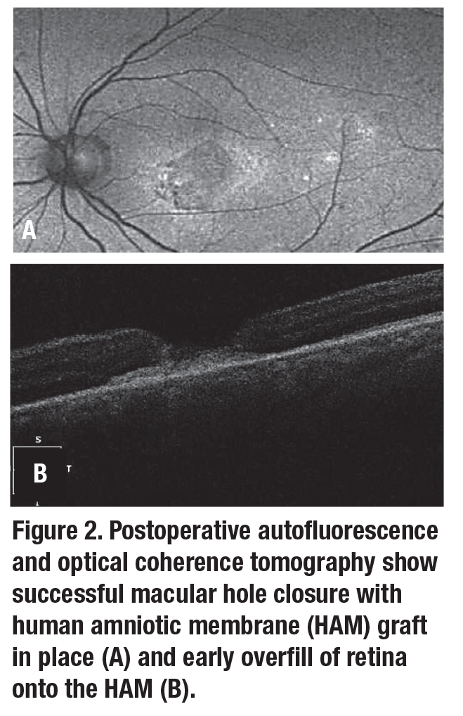
Optical coherence tomography and autofluorescence one week postoperatively demonstrated successful hole closure with maintained orientation of the HAM graft (Figure 2) and no signs of excessive intraocular inflammation.
Dr. Rizzo has reported follow-up beyond one year and hypothesized that secreted growth factors may stimulate retinal repair. This technique shows promise as an alternative for surgical closure of large, traumatic and/or recurrent macular holes.
Drs. Lau and Smith demonstrate closure of a large traumatic hole with human amniotic membrane graft.
REFERENCE
1. Rizzo S, Caporossi T, Tartaro R, et al. A human amniotic membrane plug to promote retinal breaks repair and recurrent macular hole closure. Retina. 2018;00:1-9.


