“If you were around in 1999, you remember retina specialists who said OCT didn’t give them any information that they didn’t already know,” Dr. Huang said last month while addressing the Ophthalmology Innovation Summit at the American Society of Retina Specialists. “Today, these people are using OCT everyday.”
Dr. Huang was a graduate assistant in Dr. Fujimoto’s engineering lab at Massachusetts Institute of Technology in 1991 when they co-invented OCT. Drs. Huang and Fujimoto are co-editors of a special issue of the journal Investigative Ophthalmology & Visual Science commemorating the 25th anniversary of their invention.1 More than 70 authors submitted papers on OCT for the issue.
What they started has now become a $1 billion worldwide industry that accounts for more than 2,500 jobs and more than 30 million OCT images that physicians capture each year.1
Today, Dr. Huang is a professor of ophthalmology and biomedical engineering at Casey Eye Institute at Oregon Health & Science University, and leads the Center for Ophthalmic Optics and Lasers Lab—the COOL Lab—at the institute. This year, as OCT commemorates its silver anniversary, Dr. Huang and his colleagues are more focused on the next 25 years of OCT. In an interview during OIS@ASRS, he provided insight into what’s next for OCT.
Even Faster Swept-Source OCT
While Dr. Huang doesn’t believe OCT will be the beneficiary of infinite improvements, he does see the next phase in OCT development: much higher-speed, swept-source OCT. “That would improve the speed compared to current commercial systems, which run between 70 and 100 kHz; there’s another factor of 10 by which that can improve,” he says. “Eventually, the volume will be large enough that you can have OCT on a chip so that doctors can scan many beams and have it really compact and economic.”
 |
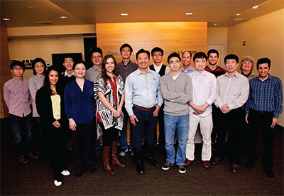
| David Huang, MD, PhD, (center in light blue shirt) with members of the Center for Ophthalmic Optics and Lasers Lab at Oregon Health & Science University Casey Eye Institute. |
The Need for Speed
Dr. Huang considers the development of OCT angiography the most significant advance for the technology in the past five years. “It’s going to really develop,” he says. “We’re going to learn how to interpret the images together with regular structural OCT: They’re really synergistic; you get both from one scan anyway. It’s not really a different data set; it’s just different ways to process the same data.”
Increasing the speed of OCT is vital for improving angioscans and widefield scanning, Dr. Huang says, because the devices are so sensitive to motion they require volumetric scanning and skilled retinal photographers who can interact with patients in ways that minimize motion artifacts.
“These will eventually make photography a less-needed skill, make fluorescein angiography less needed and probably replace some function of the scanning laser ophthalmoscope systems as well in terms of widefield imaging,” Dr. Huang says. “This will take several years to play out.”
Further into the future, perhaps in 10 years or so, he sees greater use of intraoperative OCT by retina specialists.
Incubating in the COOL Lab
Meanwhile in Oregon, Dr. Huang and his colleagues at the COOL Lab are investigating greater applications of OCT beyond the retina—namely anterior segment OCT to measure corneal topography and epithelial mass to guide selection of intraocular lenses, perform phototherapeutic keratectomy and diagnose and manage keratoconus.
There’s also what Dr. Huang calls the “big glaucoma project.” He adds, “OCT angiography is a big part of that, but even within conventional-structure imaging there are a lot of advances that will continue to come out of this translational research, meshing the anatomy better and sorting out the hallmarks of glaucoma at a finer level.”
The team is also working on novel contrast imaging. “We’re looking at a nanoparticles contrast agent,” he says. “That could be promising to be able to label cells and molecules with OCT imaging. That’s always been a deficiency of OCT compared to scanning laser ophthalmoscopy and ophthalmoscopic fluorescein imaging.”
Oximetry is another area where the COOL Lab investigators are taking OCT. That involves using spectroscopic contrast to measure oxygen levels in tissues. “That fits together with the angiography part as well,“ Dr. Huang says.
“I’ve worked on this for 25 years,” Dr. Huang says. “I could probably work on this for another 25 years. I never run out of things to do.” RS
REFERENCE
1. Fujimoto J, Huang D. Forward: 25 Years of optical coherence tomography. Invest Ophthalmol Vis Sci. 2016;57:OCTi-OCTii.


