March / April 2019
Features
Can RETINAL IMAGING predict progression to DME?
The role of optical coherence tomography, OCT angiography and ultra-widefield fluorescein angiography in identifying risk factors.
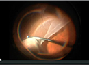
Repair of RD associated with GIANT RETINAL TEAR
Pay attention to key steps of vitrectomy to increase surgical success.

The promise of PREDICTIVE ANALYTICS in DR
Vision-threatening diabetic retinopathy is one area where risk calculators can help build better predictive models.
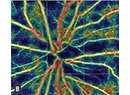
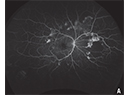
What OCT-A reveals about MACULAR PERFUSION
It may provide clues of intraocular pressure-related repetitive stress injuries.
Departments
Clinical Trial Closeup
Wet AMD gene therapy shows promise
Phase I/IIa results of RGX-314 confirm protein uptake over months.
Editor's Page
Innovation Insight
Can patients take OCT home with them?
Early work with home-based optical coherence tomography shows patients can use it, but how good are the images?
News
North of the Border
What PIVOT tells us about RRD repair
Trial authors comment on findings about the effectiveness of pneumatic retinopexy vs. pars plana vitrectomy for rhegmatogenous retinal detachment.
Retina CEO
PE: Coming soon to your neighborhood?
Private equity is here to stay, but here’s how to tell if it’s a fit, even if you’ve already jumped in.
Surgical Pearl Video
Minimally invasive IOL deposit removal
This surgical approach to introacular lens opacities utilizes a flex loop.
Current Issue

Continuing Medical Education

Additional Publications

Earlier Treatment Can Matter for Macular Edema Following Retinal Vein Occlusion
Sponsored by Regeneron
In pivotal trials, Eylea demonstrated its efficacy as a powerful treatment option for macular edema following RVO.
In pivotal trials, Eylea demonstrated its efficacy as a powerful treatment option for macular edema following RVO.
Treating Chronic Inflammation Associated With Uveitis Affecting the Posterior Segment in a Retina Setting with YUTIQ
GA: Recognizing the Burden
Treatment of Proliferative Diabetic Retinopathy
Improving Wet AMD Outcomes With a Fellow Eye Strategy
Eyepoint Yutiq: A Discussion Among Experts on the Treatment of Uveitis Affecting the Posterior Segment
